Age related macular degeneration
There are mainly two types of ARMD :
Dry AMD
Dry ARMD: where the pigment cells of the macula die and patients experience a gradual decline in central vision over many years. In late stages of this type of ARMD, the central part of vision is missing completely and patients are unable to read unless a strong magnifier is used. The Dry type is the most common form of ARMD (~90%)

Wet ARMD
Wet ARMD : in this type of ARMD, new vessels grow under the retina and leak fluid. They also tend to bleed and eventually scar. This sequence of events is what makes the central vision poor. This form of ARMD is less common but more aggressive as it can affect central vision within a few weeks. Patients experience sudden onset of distorted vision and straight lines may become wavy or bent.

ARMD will never lead to complete blindness as only is the central part of the vision is affected (macula).The rest of the retina still works normally and the peripheral vision is maintained.
Any patient over 60 years old who experiences difficulties with central vision or notices a sudden onset of distorted central vision should be examined and ruled out of ARMD.
Risk factors
The most important risk factor for ARMD is old age (>65years). Other risk factors that have been identified are:
- genes
- smoking
- poor diet
- high blood pressure
- cardiovascular risk factors
- UV light
- lack of exercise

Prevention
A healthy well balanced diet with plenty of coloured vegetables is highly recommended to prevent ARMD. It is absolutely crucial to stop smoking. Regular exercise and control of hypertension are also important as is the use of blue-blocking (i.e. brown / orange type) sunglasses to prevent UV light exposure.

Work-up and diagnosis of ARMD
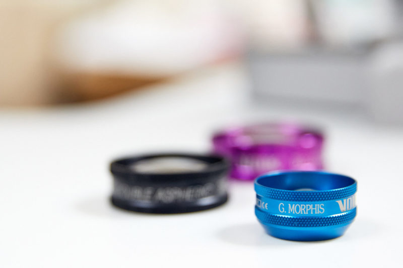
A detailed ophthalmic examination is required by a eye specialist. The examination of the retina and macula with special lenses is essential to look for changes suspicious of ARMD.
Optical Coherence Tomography (OCT) is sophisticated eye equipment which is also used to scan the back of the eye and yield information regarding the status of the macula which is sometimes not visible with the naked eye. If the suspicion of wet ARMD is high and the symptoms suggestive then a Fundus Fluorescein Angiogram (FFA) is performed to confirm the diagnosis.
The FFA is a dynamic test where a dye called fluorescein is injected in one of the veins in the back of the hand. The dye circulates in the retinal vessels and then sequential pictures are taken using a camera with special filters. The pattern of leaking vessels confirms the diagnosis of wet ARMD.
Treatment
Once the diagnosis of wet ARMD is confirmed treatment is available for patients that have reasonable levels of vision according to specific guidelines. Treatment is in the form of anti-vascular endothelial growth factors (antiVEGFs) either ranibizumab (Lucentis) or aflibercept (Eylea) that are injected in the eye.
The aim of the treatment is to maintain the level of vision the patients have or even slightly improve it. Therefore it is essential that wet ARMD is picked up early and treatment started with no delay. Lucentis or Eylea intraocular injection treatment is NOT a one off treatment. Patients need to have 3 initial injections 4 weeks apart to complete the loading phase. Thereafter close follow-ups are required with monthly OCT scans to determine the further need for injections. Most patients have 6-7 injections/year and this can continue for more than 2 years depending on the response. The treatment regimes followed for Eylea are slightly different than Lucentis with patients having fixed injection treatments during the first year.
What happens on the day of the injection?
Most patients are concerned with the idea of having an injection into their eye.
George Morphis will clean with iodine and apply anaesthetic to the eye to be injected. To minimise the risk of infection, he will also cover the face with a sterile drape and put a small clip in to keep the eye open. The volume of the injection is tiny, about 1/20 of a millilitre. The injection should feel like a pressure and not be painful.
On the day of the treatment, if you experience any discomfort, paracetamol alone is usually sufficient for any minor discomfort. The eye may also look red but this is expected to go away within the next few days. You may see floaters or spots which will also go away within 24-48 hours. In the extremely rare event of you experiencing any increasing/ excruciating eye pain, blurring or loss of vision, you should seek immediate medical advice or attend eye casualty; very rarely injections can cause a serious infection inside the eye called endophthalmitis. George Morphis will provide you with a telephone number to contact him personally in the event of any concern.
How effective is the treatment ?
Most patients find that their vision remains stable and around one third appreciate a noticeable improvement.
What happens if the eye is not treated?
Eyes with confirmed wet ARMD will gradually get worse in terms of central vision if left untreated. Leaking blood vessels will scar and detailed vision will be compromised. It is essential that patients keep their monthly appointments to determine the need for further treatments if necessary.
